Confocal Microscopy
Advertisement
Carl Zeiss Vision AxioVision Viewer v.3.0
AxioVision allows to visualize and present your images in several dimensions. The functionality of this imaging toolbox expands constantly with a wide range of different modules that are tailored to specific applications or microscope accessories.
Advertisement
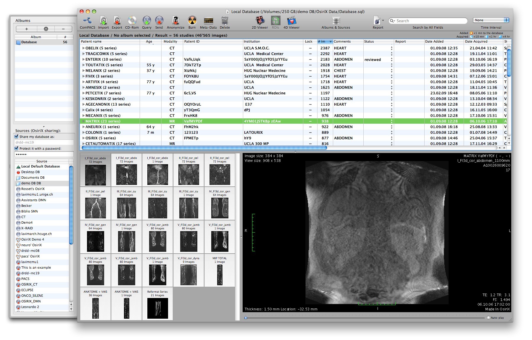
OsiriX App v.3.9.2002
OsiriX DICOM ViewerOsiriX is an image processing software dedicated to DICOM images (".
Visual3D v.1.2 Build 12
Generate a 3D visualization of confocal and wide field fluorescence microscopy images. Visual3D uses OpenGL textures to generate a 3D visualization of confocal and wide field fluorescence microscopy images.
BioView v.1.1.18
bioView was designed to be an open source and cross-platform application intended for biologists to visualize EM, Confocal, etc.
ImageSurfer v.1 24
ImageSurfer is free 3D imaging software to visualize and analyze multi-channel volumes. Main Features: - Processing: 3D filters to improve signal-to-noise ratio.
BleachingCorrection v.20. 12. 2004
The bleach correction macro for ImageJ corrects for bleaching or intensity fluctuations by normalizing the images of a stack to the same mean intensity. This method only works well if the mean intensity is not altered substantially (e.g.
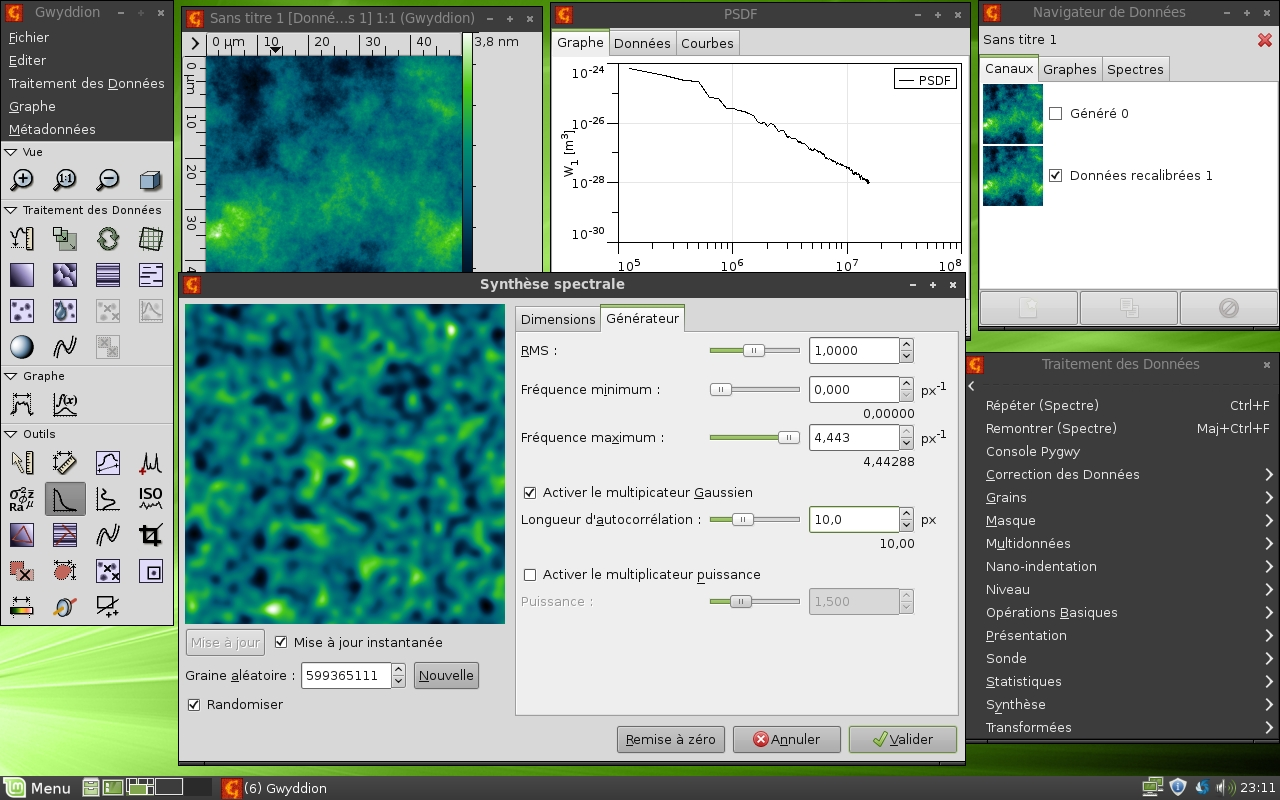
Gwyddion v.2.23
Gwyddion is a modular program for SPM (scanning probe microscopy) data visualization and analysis.
GXSM for Windows v.2.0 Alpha
GXSM - Gnome X Scanning Microscopy - is a strong graphical interface for any kind of 2D and 3D (multilayered 2D mode) data acquisition methods, especially designed for scanning probe microscopy (SPM).
Deconvolve v.2.1 Build 10
Deconvolve is a Windows software for remote batch restorations of 3D/2D confocal and widefield fluorescence images using Huygens2 from SVI on a local or remote computer.
Neuron Analysis v.Beta
Tool for analyzing neuron pictures. Image Analysis program designed specifically for the analysis of images of neurons acquired with a confocal microscope or a fluroscence microscope.